Hello Dear FLUKA experts
I’m comparing dose distributions between FLUKA simulation and a clinical RTDOSE DICOM file.
Although I’m using the same voxel dimensions and binning (XYZ) as the RTDOSE, the FLUKA-calculated dose appears spatially shifted from the RTDOSE map.
Here are more details:
Voxel geometry is imported from DICOM CT.
USRBIN is used for scoring dose (DOSE).
The bin sizes, number of bins, and origin match those from the RTDOSE.
RTDOSE and CT data are aligned in the treatment planning system (TPS).
My questions:
Is there a known issue or best practice for ensuring alignment between FLUKA dose output and RTDOSE distribution?
When converting DICOM to FLUKA input using DICOM2FLUKA or Voxel tools, is the isocenter automatically placed into ROT-DEFI or does it have to be manually inserted in cards like BEAMPOS CARD?
Any clarification on isocenter handling and coordinate systems would be really helpful.
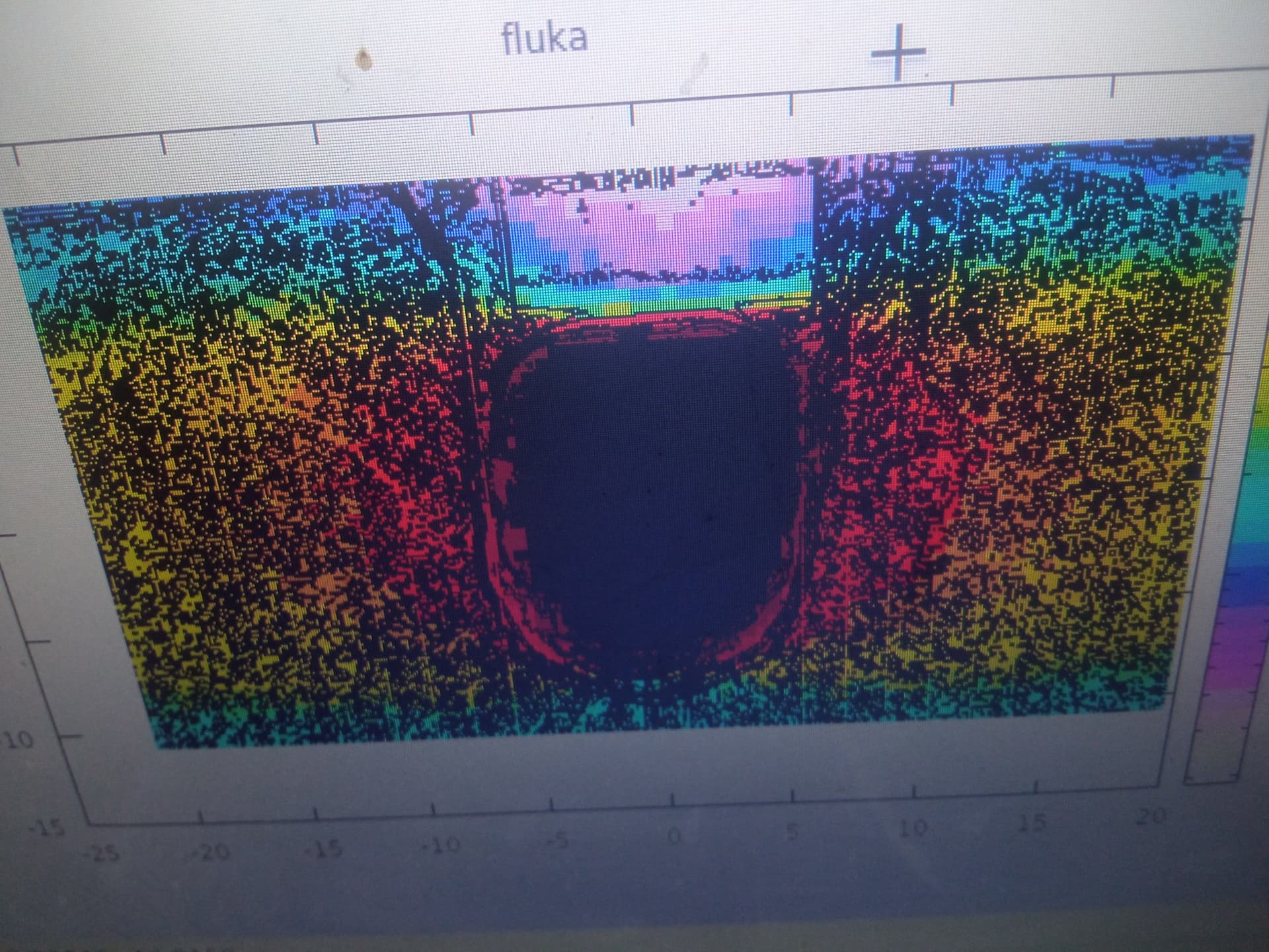
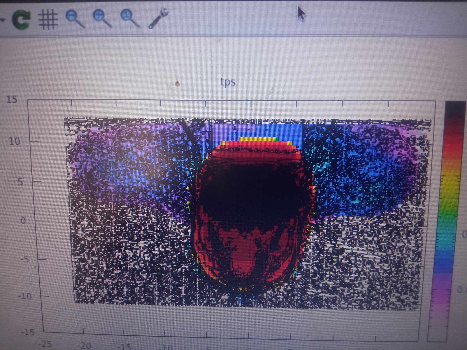
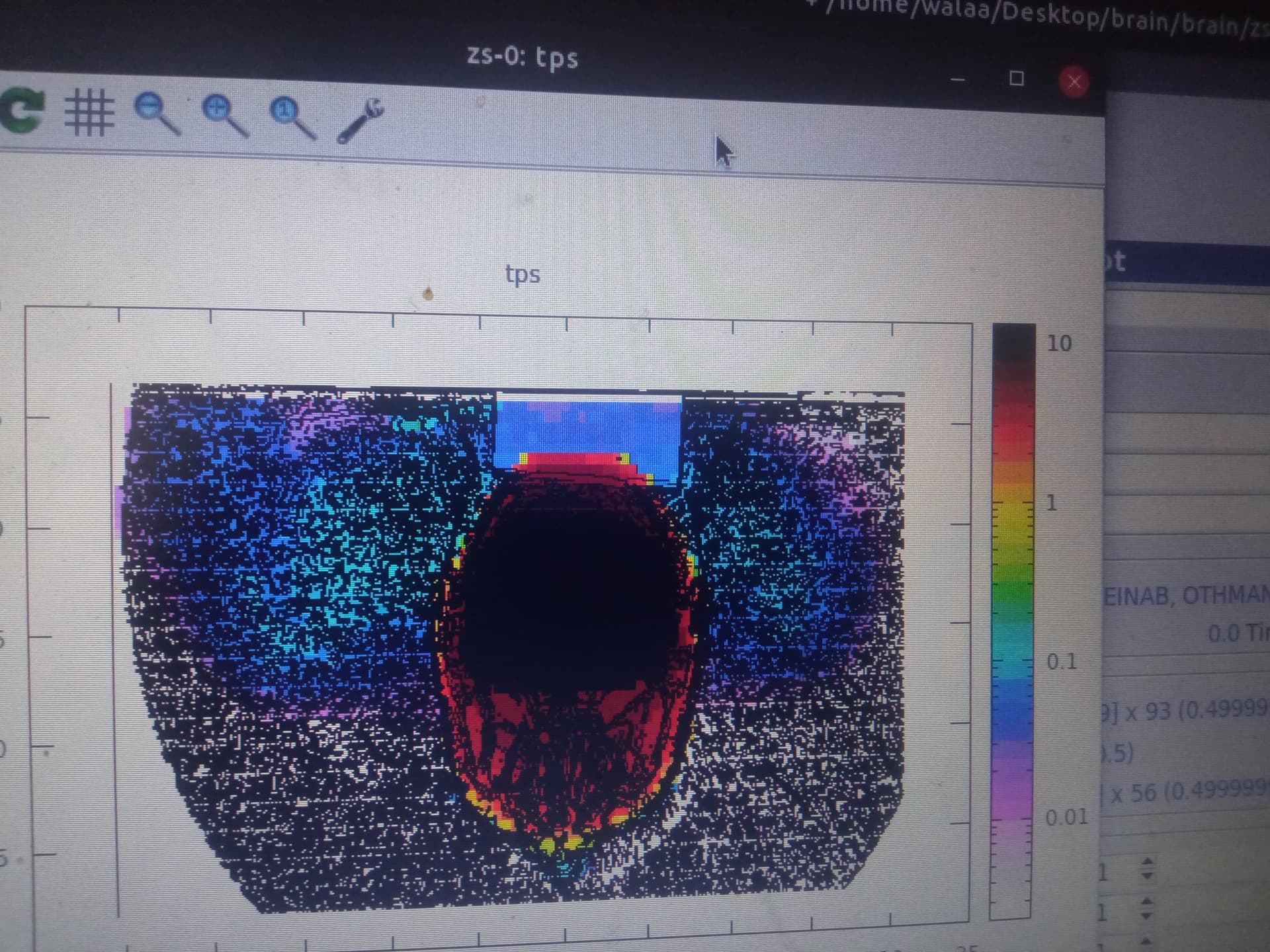
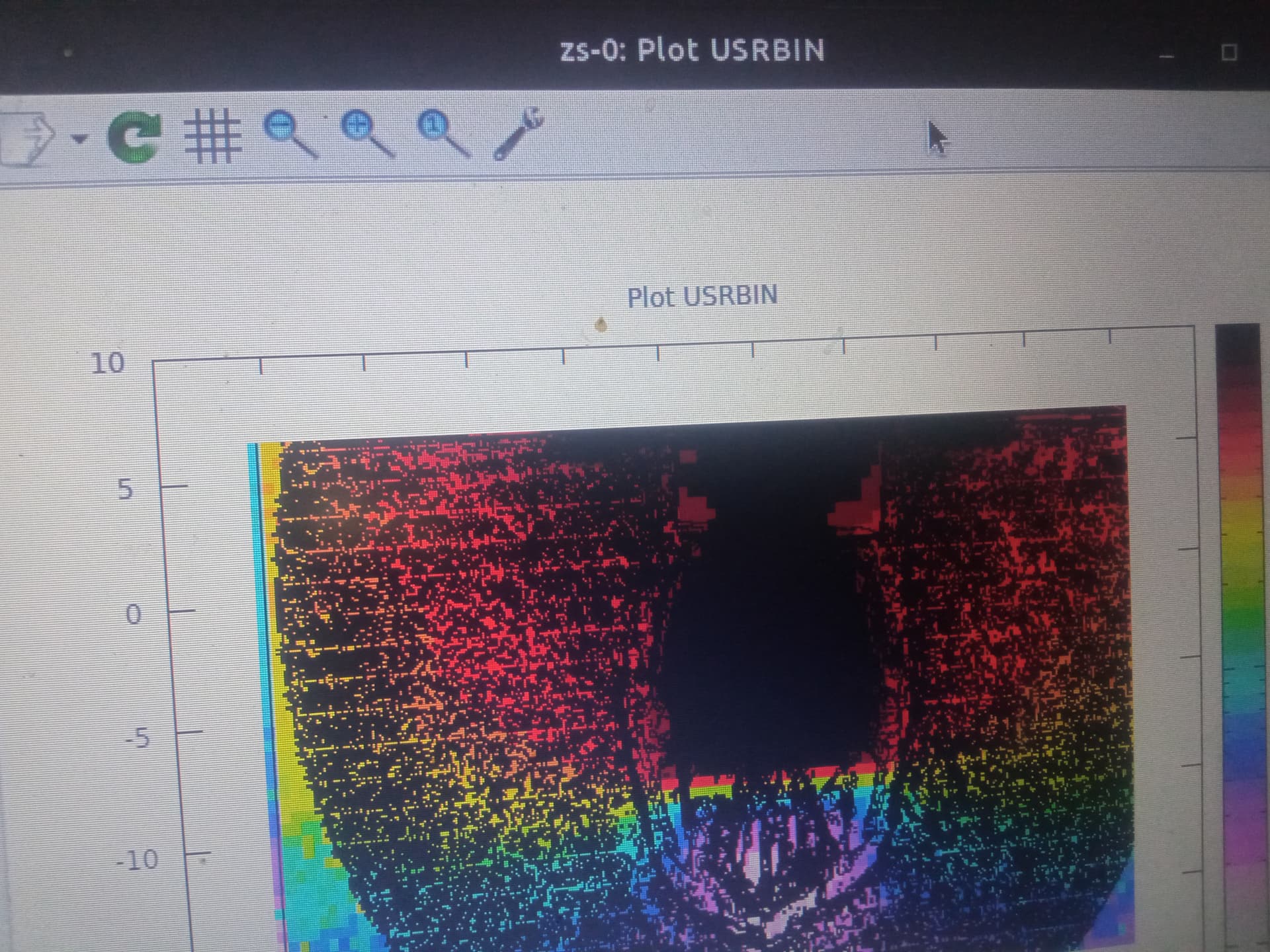
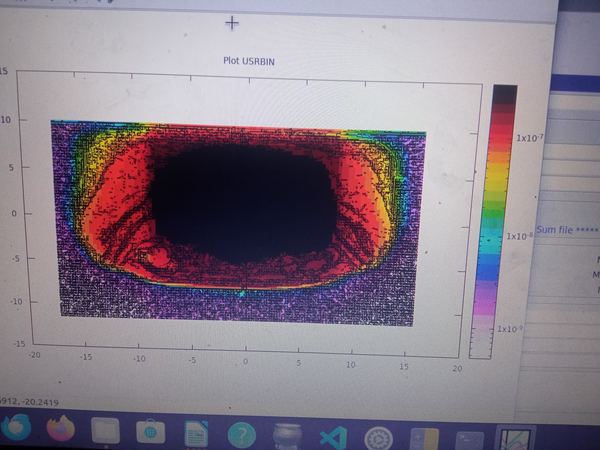
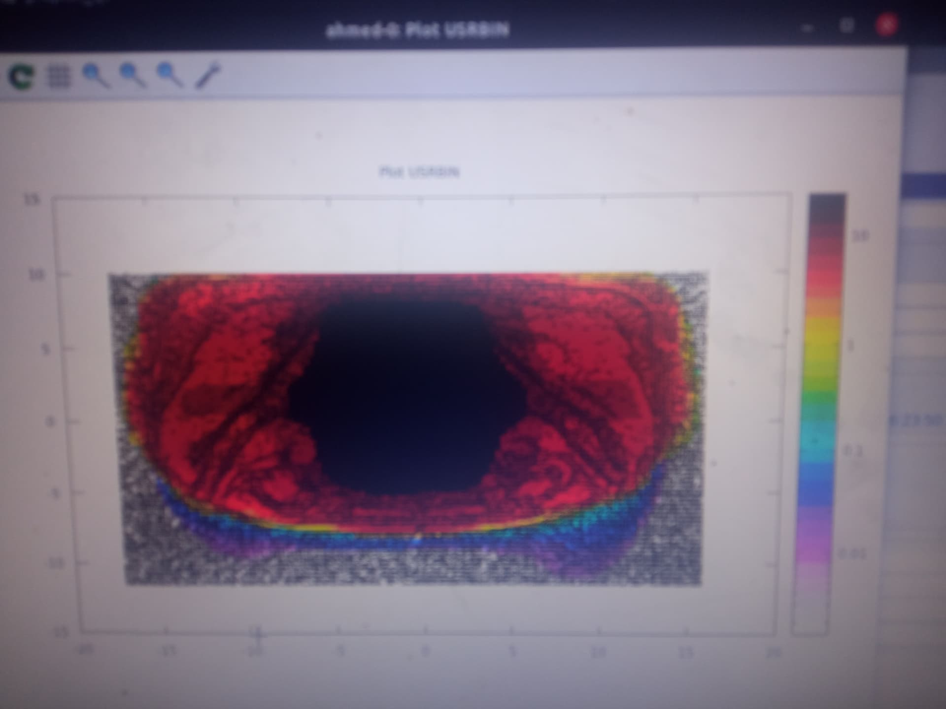
In these images fluka dose closed eyes this makes dose of eye is very high
RTDOSE blocked eyes by multileaf collimators , I saw ISOCENTER FLUKA inserted It above the brain is so that the dose shifted to above?
but in this case FLUKA field dose is very larger than RTDOSE field .why ?
are the mltileaf collimators blocked the field in RTDOSE if yes how I can insert them in FLUKA ?
Thanks!
Hi
Dear@vasilis @FLUKA_Admin
I would be grateful if you could take a look or guide me on this
Just following up on this issue. I’m still seeing the dose distribution mismatch and would like to understand whether it’s related to isocenter placement or voxel origin
Thanks in advance
I’m comparing dose distributions between FLUKA simulation and a clinical RTDOSE DICOM file from treatment planning system. the fluka dose apears spatially shifted from the expected location.
Although I’m using the same voxel dimensions and binning (XYZ) as the RTDOSE, the FLUKA-calculated dose appears spatially shifted from the RTDOSE map.
Here are more details:
Voxel geometry is imported from DICOM CT.
USRBIN is used for scoring dose (DOSE). The same dimensions of the USRBIN of the RTDOSE . The bin sizes, number of bins, and origin match those from the RTDOSE.
RTDOSE and CT data are aligned in the treatment planning system (TPS).
My questions:
Is there a known issue or best practice for ensuring alignment between FLUKA dose output and RTDOSE distribution?
When converting DICOM to FLUKA input using DICOM2FLUKA or Voxel tools, is the isocenter automatically placed into ROT-DEFI or does it have to be manually inserted in cards like VOXEL or BEAMPOS card?
Any clarification on isocenter handling and coordinate systems would be really helpful. Thanks!
Here the link of the original issue.
normally the flair tools should take care for the proper alignment and place everything in the isocenter. However the DICOM format is so versatile that allows multiple ways of describing the information, so it is not unlikely that flair didn’t manage to get the proper information to align the various information.
The only way to check would be if you could give us access (privately) to the dicom data.
Dear@vasilis
Thank you for your support and willingness to help.
Unfortunately, I’m not allowed to share DICOM files due to my university’s data protection policy. I sincerely apologize for this and hope for your understanding.
I can share:
Screenshots showing the voxel size, origin, and RTDOSE spacing.
The FLUKA input cards related to geometry (VOXEL, USRBIN, ROT-DEFI) if helpful.
Any specific information you request that can be safely extracted from the DICOM using Python (e.g., voxel dimensions, image position/pixel spacing from the RTDOSE or RTPLAN).
Please let me know what exact data you’d find most useful, and I’ll provide it accordingly.
Thanks.
Please can you tell me what you need from the dicom file to help me know the reason of the mismatch.
the input file or specific information that can be safely extracted from the DICOM using Python.
Thanks.
Unfortunately it will be extremely complicated to debug/understand like that. Potentially if you could provide the dump of the dicoms as python reads them could help. (and send privately)
You can do the following with python for at least one file for each modality: CT, RTDOSE, RTPLAN
<dicom.file> with the dicom file CT, RTDOSE, RTPLAN
Open each text file in an editor and remove any sensitive information like: Patient name, birth date, etc…
By default it will write only the metadata of the dicom, not the image data.
Dear@vasilis
Thank you very much for your continuous support.
Can you tell me how i can contact with you (privately) to sent the files that you requested directory and security.
Thank you in advance for your cooperation.