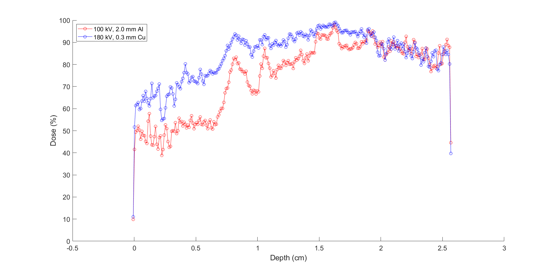
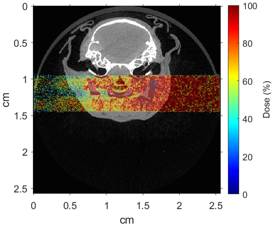
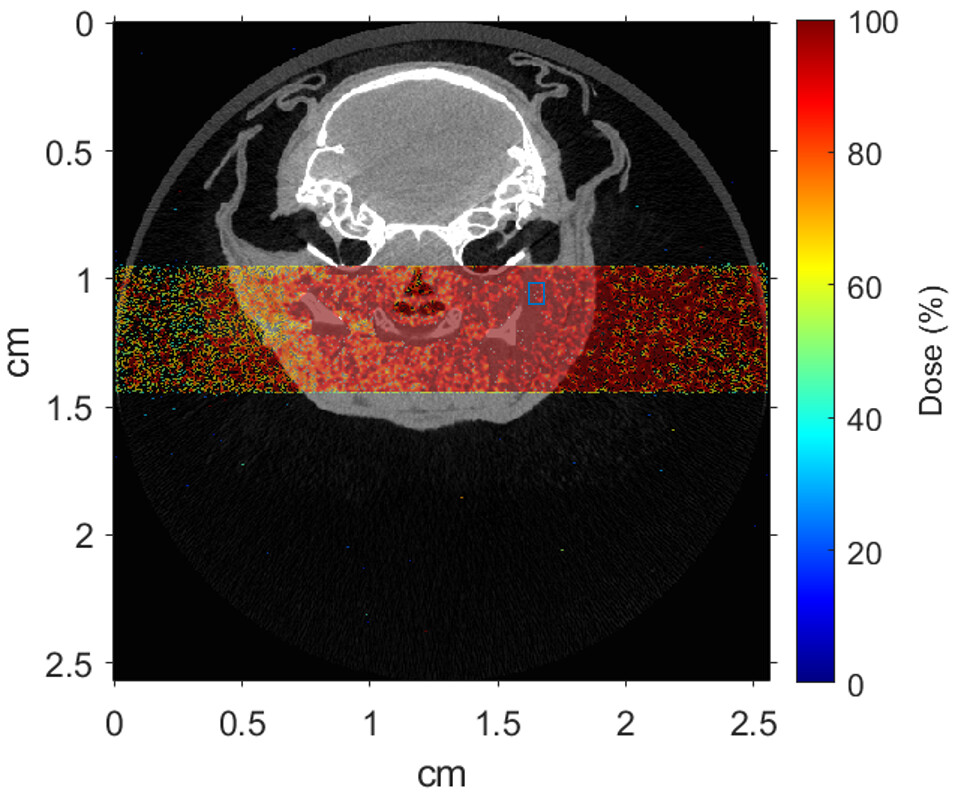
I have successfully imported DICOM CT images (of a mouse) and am trying to score dose deposition from a 5 mm x 5 mm rectangular X-ray beam of 100 kV energy given sagitally to the neck of the mouse. The energy spectrum, which is sampled from the file ‘Spec100f’, is “pre-filtered” (to save computation time) by 2.0 mm Al. However, importing the generated ASCII file of the .bnn scoring detector into Matlab, I am getting high dose depositions (allegedly) to air before the X-rays hit the skin. I have included a color wash plot of a single CT slice (central to the beam axis) showing the normalized dose. The dose map is normalized relative to a pre-selected ROI as pointed in the color wash plot. Furthermore, to examine this, I changed the beam setting to 180 kV X-rays filtered by 0.3 mm Cu (harder filtration) and am still getting the same high relative dose deposition to air (here I have plotted dose profile along the beam axis in the same CT slice for the two X-ray settings). Why am I getting such high doses to air? Is there something wrong with my voxel cage? I have checked the re-scaled values of the air voxels in the CT slice in FLUKA, which is in the range of [-1000, -800].
I can confirm that the FLUKA dose maps are read correctly as I superimposed the generated .bnn dose file (by adding a Usrbin) on the 3D layer in Geoviewer and verified it with current axial slice (as posted). Does that mean something is off with the normalization?
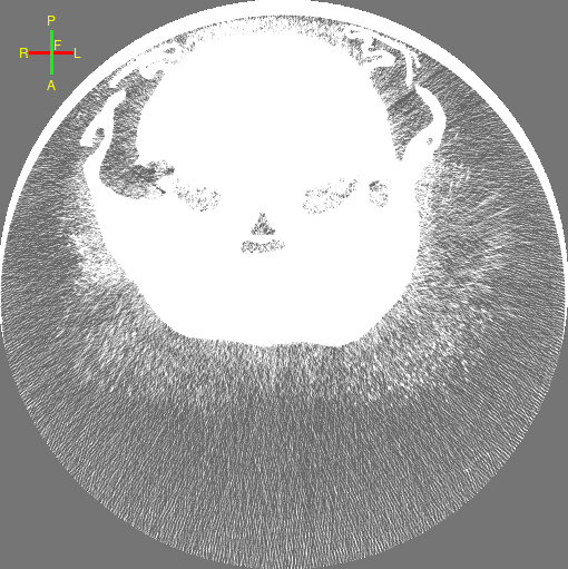
This way you can see the hair of the mouse (and maybe some distortion of the CT image ?). Since these regions are not clear black as air should be, their voxel regions are filled with dense material, which leads to dose deposition there.
You may try to edit the material of these lower density regions on the page for voxel generation (See: “Voxel Generation” at Flair manual, but this could lead to changes at some internal organs as well.