Scoring secondary particles produced by carbon ion beam simulation in a water target

Versions

FLUKA:version: 4-5.2
Flair:3.4-5.2

Description

Hello team,

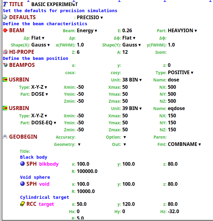
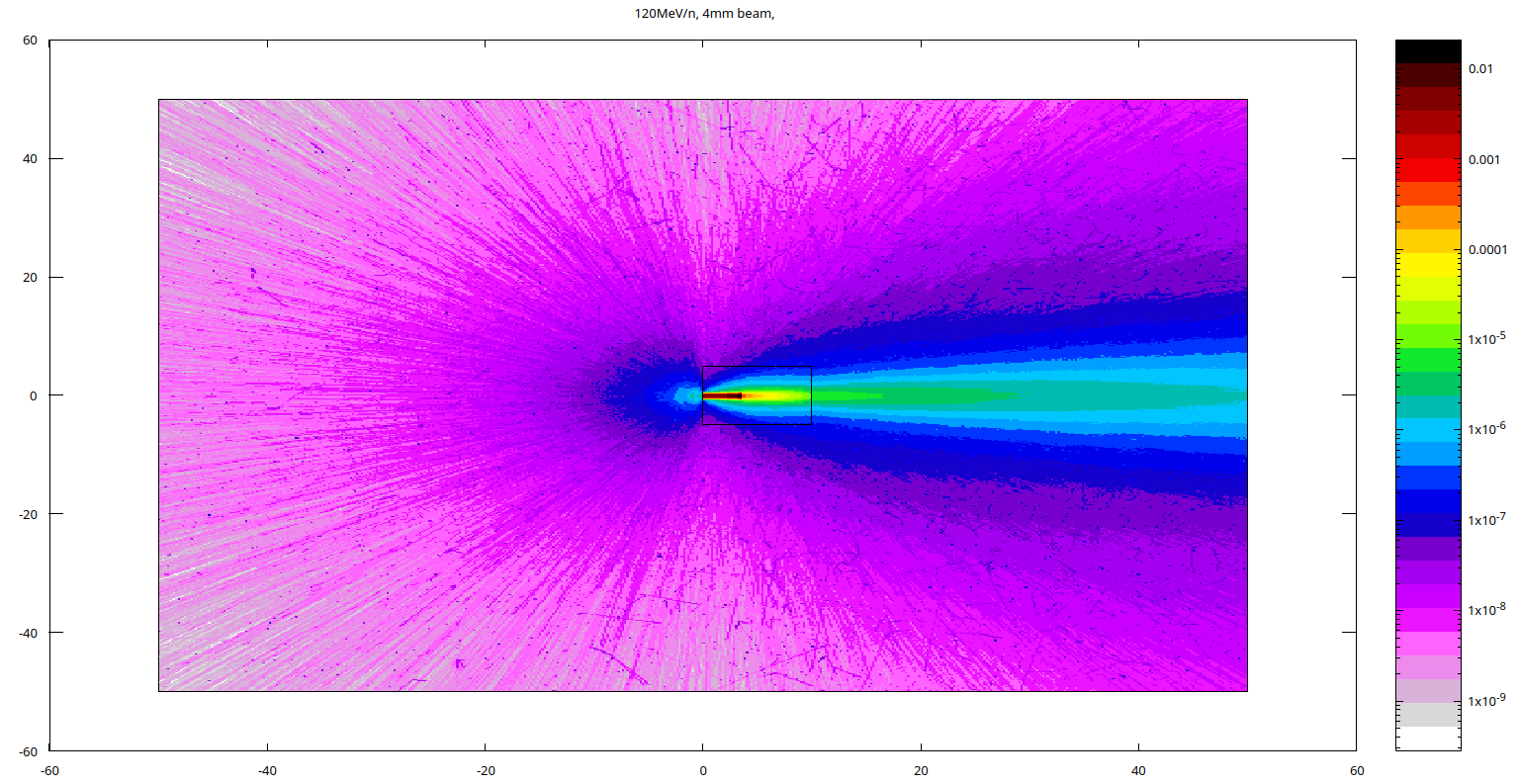
  1. I simulated a carbon ion beam through a water target and obtained this color distribution in the picture attached. However, my professor says the image shows unnecessary wide color band and that its not correct. please look at the input and advise.
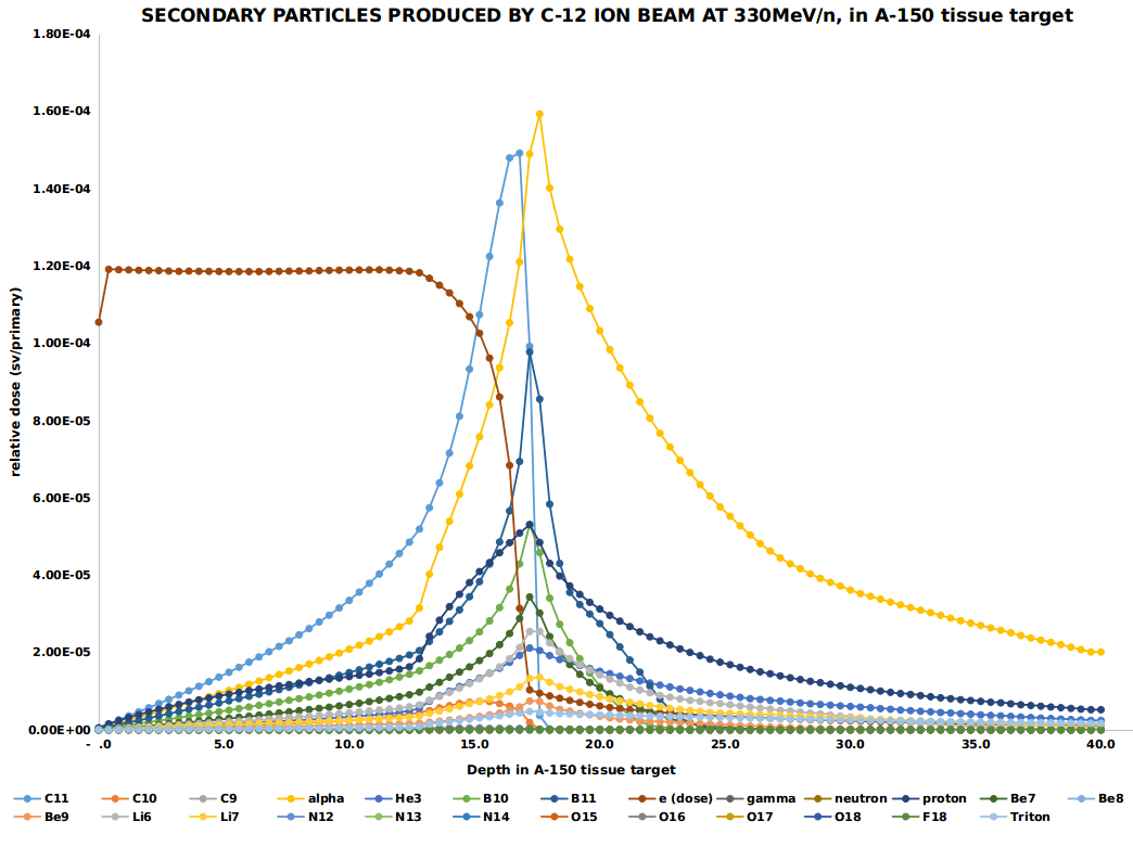
  2. I used a set up attached as input file to simulate secondary particles and I obtained the graphs also attached, which my professor still says its not correct. please advise on where the mistake is on how i set up the inputs. Thanks.
  3. I need to clarify that the above are two different inputs, one (picture of input) for only the primary beam and the other(input file) for secondary particles

Input files

Please upload al

LARGE TARGET ONONO CARBON ION BEAM DESIGN.inp (11.2 KB)

l relevant files. (FLUKA input file, Flair project file, user routines, and data files)
Dear @ononoalex,

There are a few things to mention:

  1. In the *.inp file you attach, your BEAM starts exactly at the boundary of your WALL region. It is typically advised to have your BEAM positioned within one region, and not at the boundary - though this should not impact your simulation.

  2. You have a rather simple case of a beam hitting a target - and the resulting dose distribution looks reasonable. Why do you believe/what reference do you have that:

  1. Your 260 MeV/n Carbon ions stop and have a Bragg peak energy deposition pattern inside your target, as expected. The forward shower that exits the target is 3 orders of magnitude lower than the peak inside the target, from light secondary particles that travel further. You also have particles (mostly electrons and gammas) that are generated at large scattering angles, which also exit the target - but these are low energy compared to your initial beam and hence why a minuscule (6 orders of magnitude compared to Bragg peak) dose deposition.

  2. Regarding the secondary particles - you are plotting their dose-equivalent deposition, and not their fluence. Consistent with my point above (3), only lighter fragments (particularly alphas) penetrate further in the target. Again, what is the reference to conclude that:

  1. For the future, please upload the *.inp files, not screenshots of the *.inp files (for your target study case).

Best,
Daniel

Thank you for your constructive feedback. Let me simulate the particle fluence of the secondary particles to find out the difference in the plots.